Our Investment in Your Health
At McCarroll Dental we use the latest technology to assist us in the planning and delivery of our dental services to patients. Modern Dentistry continues to evolve every year and it is our mission to research and employ the best technology to treat our patients comfortably and successfully.
Digital X-Rays
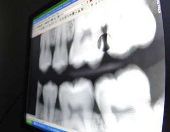 Digital X-Rays replace bulky standard X-Ray film with a small more comfortable sensor that captures the image and is then used to scan into a computer. By scanning and storing your x-rays on a computer we eliminate caustic developing solutions, lead foil and other agents used with traditional x-ray film. Also, digital x-rays allows us to pull up our films on the screen positioned next to our treatment chair and discuss with you the diagnosis of what we see and recommended treatment. Environmentally friendly, comfortable, less radiation to the patient and easily shared on the computer screen makes digital x-rays a great diagnostic solution for us and our patients.
Three-Dimensional Imaging
 Dental Cone Beam CT (CBCT) is currently at the cutting edge of Dentistry for its ability to diagnose issues and plan treatment in three dimensions instead of two by rendering volume to each scan. Our state of the art 3D imaging scanner allows us to detect things that may have alluded us on standard 2D imaging. Also, 3D imaging is a remarkable tool in the planning and placement of implants whether it be a single or multiple implants. It allows us to view how much available bone is present in the area of interest with incredible precision making implant placement safer and much more predictable for our patients.
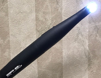
Soft-Tissue Dental Laser
 We use a diode laser in many procedures to enhance comfort and promote quick healing where gum and tissue lining treatment is necessary. The laser helps reduce and seal soft tissue with no bleeding, allowing us to quickly and comfortably complete tissue and gum therapy to promote optimum oral health for our patients. Learn more about laser dentistry in our office.
Intraoral Camera
 We employ an intraoral camera in our office to capture images of teeth, gums and surrounding tissues of the mouth in order to better communicate with our patients their existing conditions and ways to treat them. This device takes a very clear picture that shows in great detail conditions that a patient would otherwise not be able to view. For example, cracks in teeth can form around old fillings and patients may be totally unaware until we take an intraoral photograph and share it with them on the computer screen.
Our patients continually find it amazing when they see images of their teeth and exclaim “I never realized they looked like that” or “That’s amazing!”
The Wand Anesthesia System
We are very proud to offer our patients a method of anesthesia that is as comfortable as we can get it. Instead of a traditional syringe which is bulky and more cumbersome we use a device like a pen that can be gently guided to the appropriate spot and administered a 3 different flow speeds: Slow, Medium. and Fast depending upon the comfort of the patient. We have used this system for 20 years (since it began) and can’t say enough about the difference it makes for our patients.